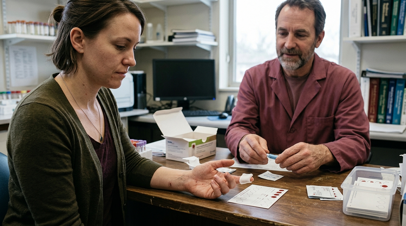

Clear insights into dried blood spot hormone testing help you evaluate finger-prick testosterone accuracy and laboratory limitations compared to standard serum blood draws.





Dried blood spot testing is a collection method that uses dried whole blood from a finger prick to analyze specific compounds in a laboratory. It is not a direct substitute for standard venous blood testing, nor does it provide an identical measurement to blood serum. Understanding what this technology can reliably measure, how it behaves under laboratory analysis, and where its limits lie is essential for interpreting any hormone panel.
This guide provides a detailed look at the science behind dried blood spot testing. It covers laboratory validation, matrix differences, the physics of blood spreading, and how clinical guidelines view these results when assessing male hormonal health.
This article is for educational and informational purposes only. It is not personal medical advice, diagnosis, or treatment. Hormone levels, laboratory results, and medical symptoms require individual assessment by a qualified healthcare professional. Never disregard professional medical advice or delay seeking it because of information read here.
Dried blood spot collection provides convenience, but it operates under different physical and biochemical principles than standard clinical testing. Keep the following evidence-based points in mind:
Dried blood spot testing involves collecting a few drops of capillary blood, usually from a simple finger prick. The blood is applied to specialized filter paper cards, allowed to air-dry thoroughly, and sent to a laboratory for analysis. Once at the facility, technicians extract the dried chemical material from the paper matrix and analyze it using techniques like liquid chromatography tandem mass spectrometry or traditional immunoassays.
This collection method was originally developed for newborn metabolic screening. Over recent decades, researchers have adapted it to measure various hormones, therapeutic drugs, and environmental markers. Because the collection paper stabilizes many molecules when dry, samples can often travel through standard postal systems without complex refrigeration. This makes remote collection possible for research studies and home health tracking.
However, the simplicity of collection often obscures the laboratory complexity. A dried spot is not a miniature vial of serum. It is an unseparated mixture of cellular components, fluid, and paper fibers. Laboratories must use rigorous chemical extraction steps to separate the target hormone from the paper before measurement can occur.
The reliability of the final
number depends heavily on every step in the sequence. These steps include the precision of the paper card, the technique used during the finger prick, the ambient humidity during drying, and the specific assay technology used by the testing facility. Learning the foundations of hormone testing protocols helps contextualize how these laboratory workflows operate.
In laboratory medicine, the term "sample matrix" refers to the entire physical and chemical environment surrounding the molecule being measured. The matrix fundamentally alters how an assay behaves. Traditional clinical testing uses venous blood, which is drawn from a vein in the arm. The clinical laboratory spins this blood down in a centrifuge to remove red blood cells, white blood cells, and clotting proteins, leaving behind clear liquid serum.
Dried blood spot testing uses whole capillary blood. Whole blood contains red blood cells, white blood cells, platelets, and water, along with dissolved proteins and electrolytes. When a laboratory measures testosterone or any other steroid in a dried spot, it is measuring the hormone across this entire cellular mixture rather than in clear serum alone.
Steroid hormones such as testosterone distribute themselves unevenly across these components. In the bloodstream, the majority of testosterone binds tightly to sex hormone-binding globulin or loosely to albumin. A small fraction remains unbound as free testosterone. Testosterone can also partition into red blood cell membranes, altering the ratio of hormone found inside cells compared to the surrounding fluid.
Because whole blood has a different chemical composition than serum, matching units on a report do not mean the biological measurements are identical. A laboratory value of 500 nanograms per deciliter reported from a dried spot extract is derived from a distinct analytical procedure. It cannot be assumed to equal 500 nanograms per deciliter from a standard venous serum draw. Converting a whole blood concentration to a serum-equivalent estimate requires rigorous, published validation studies rather than simple unit matching.
To understand these measurements clearly, three distinct concepts must remain separate:
Hematocrit represents the percentage of total blood volume made up of red blood cells. In healthy adult men, hematocrit typically ranges from forty to fifty-two percent, though it can vary based on hydration, altitude, genetics, and medications. Hematocrit directly affects blood thickness, which in turn determines how quickly a drop of blood spreads when it hits filter paper.
When blood has a high hematocrit, it is more viscous. A viscous drop spreads slowly and forms a smaller, thicker spot on the collection paper. When blood has a low hematocrit, it is thinner and spreads widely, creating a larger, thinner spot. This physical spreading behavior introduces a significant variable known in clinical chemistry as the hematocrit effect.
Most standard laboratory workflows do not analyze an entire dried blood spot. Instead, an automated mechanical punch cuts out a small, uniform circular disc from the center of the spot, such as a three-millimeter or six-millimeter circle. The laboratory assay assumes that every disc contains an identical, fixed volume of blood.
If a patient has a high hematocrit, a three-millimeter punch will contain more red blood cells and more total blood volume than the laboratory calculation assumes. If the patient has a low hematocrit, the same punch contains less blood. Method validation studies published in analytical chemistry journals show that hematocrit differences can create an analytical bias of up to twenty percent in reported hormone concentrations.
Spot volume adds another physical challenge. If a user deposits a small drop of ten microliters versus a large drop of sixty microliters, the rate of absorption and the density of the dried ring can vary. Research evaluating dried blood spot testosterone protocols indicates that spot volume alone can introduce another twenty percent variance if the collection is not strictly controlled.
Some modern techniques attempt to bypass this problem by analyzing the entire dried spot rather than a small punch. Other methods require a separate hematocrit measurement to mathematically correct the final value. However, measuring hematocrit accurately requires a traditional blood draw, which reduces the practical convenience of finger-prick testing.
Evaluating the reliability of dried blood spot testosterone testing requires separating laboratory analytical validity from clinical diagnostic performance. A test can be highly repeatable in a controlled research facility without necessarily being suitable for diagnosing medical conditions in a clinic.
Recent validation research using liquid chromatography tandem mass spectrometry has shown that dried blood spot assays can achieve impressive technical specifications. In controlled laboratory environments, modern assays report linear measurement across a broad range from 0.1 to 100 nanograms per milliliter. Researchers have documented lower limits of quantification as low as 0.086 nanograms per milliliter, with intra-day and inter-day precision errors held under ten percent.
These technical figures confirm that a mass spectrometer can detect and measure testosterone molecules extracted from paper with high repeatability. Furthermore, stability studies demonstrate that steroid hormones dried on quality filter paper remain stable for several weeks at ambient room temperature or under refrigeration.
Problems arise when comparing these laboratory measurements directly to clinical venous samples. In a 2024 validation trial comparing corrected dried blood spot testosterone to venous serum in twenty adult subjects, researchers observed a strong statistical correlation coefficient above 0.98. While this high correlation proves that the two measurements move in the same direction, correlation is not mathematical agreement.
A statistical correlation does not establish whether a dried blood spot test and a serum test yield identical numbers at a specific diagnostic boundary. The validation study did not report Bland-Altman numerical limits of agreement, which clinicians use to determine if two diagnostic tools can be used interchangeably. Additionally, the study used an immunoassay for the serum comparison rather than matching mass spectrometry techniques, introducing another variable into the comparison.
Collection conditions in real-world settings also show wider variation than research trials. In study settings where volunteers collected their own samples at home, the correlation factor dropped compared to samples collected on-site by trained personnel. This highlights how user technique, drying conditions, and card handling affect final data quality before the sample ever reaches a machine.
Commercial testing services frequently offer multi-hormone panels on a single dried blood spot card. These panels promise to measure total testosterone, estradiol, progesterone, DHEA, cortisol, and other steroids from one finger prick. However, validating a method for one hormone does not validate it for every compound on the card.
Steroid hormones differ fundamentally in their chemical structures, molecular weights, and circulating concentrations. Testosterone circulates in adult men at nanogram-per-milliliter concentrations, whereas estradiol circulates at much lower picogram-per-milliliter concentrations. Measuring a compound present in minute quantities requires far greater analytical sensitivity, leaving less room for sample-spreading errors.
Each steroid also partitions differently between red blood cells and plasma water. In a peer-reviewed investigation comparing multiple steroid concentrations measured in dried blood spots against matching serum, researchers found dramatic differences across analytes:
These findings show that each hormone interacts uniquely with the paper matrix and extraction solvents. A laboratory cannot apply a single universal correction factor across an entire panel of hormones. Every individual biomarker must undergo independent validation, including tests for recovery efficiency, chemical interference, and matrix effects.
Differences also appear when comparing male and female physiology. Historical research evaluating gonadal hormones in dried blood spots found that blood spot values accounted for roughly eighty-eight percent of the variance in female serum samples, but only forty-six percent of the variance in male samples. Because hormone baseline concentrations and binding protein profiles differ by sex, assay behavior changes accordingly.
When reviewing a multi-hormone panel, evidence must be evaluated compound by compound. High accuracy for one marker provides no guarantee that adjacent markers on the same report meet the same standard of precision.
Laboratory measurements never exist in a vacuum. In clinical medicine, biomarker data must be evaluated alongside clinical history, physical symptoms, and established diagnostic guidelines. The Endocrine Society provides clear, evidence-based recommendations for assessing male hypogonadism and interpreting testosterone fundamentals.
According to clinical practice guidelines, the diagnosis of androgen deficiency requires two essential components:
The requirement for morning fasting draws is based on human circadian biology. Testosterone levels peak in the early morning hours and decline throughout the afternoon in young and middle-aged men. Food intake can also temporarily lower circulating testosterone by twenty to thirty percent.
Because standard clinical thresholds (such as 300 nanograms per deciliter) were established using morning venous blood draws, they cannot be applied directly to a capillary dried blood spot collection. If a dried blood spot test shows a borderline result, it cannot confirm a medical diagnosis on its own. Clinicians require standard venous draws to determine whether hormone levels genuinely fall below reference intervals.
Furthermore, total testosterone tells only part of the biological story. In patients with conditions that alter sex hormone-binding globulin, such as obesity, diabetes, liver disease, or aging, total testosterone can appear misleadingly low or high. In these cases, guidelines recommend measuring or calculating free testosterone using equilibrium dialysis or validated formulas based on total testosterone, binding globulin, and albumin.
Current dried blood spot methods rarely measure sex hormone-binding globulin and free testosterone with the precision required for complex clinical evaluations. Understanding the details of evaluating low testosterone requires looking at the entire hormonal axis rather than relying on an isolated finger-prick value.
To understand what dried blood spot reports show, it helps to examine how individual biomarkers perform in capillary whole blood versus standard venous serum.
Total testosterone represents the combined total of bound and unbound hormone circulating in the blood.
Free testosterone is the unbound fraction of the hormone, representing roughly one to two percent of total circulating testosterone. It is biologically active and able to diffuse into tissues.
Sex hormone-binding globulin is a glycoprotein produced by the liver that binds tightly to testosterone and estradiol, regulating their transport and biological availability.
These pituitary gonadotropins signal the testes to produce testosterone and sperm. They are essential for differentiating primary testicular failure from secondary pituitary or hypothalamic dysfunction.
You can learn more about how these hormones interact by reading our dedicated resource on laboratory testing and biomarkers.
To see how these laboratory principles apply in everyday situations, consider four common practical scenarios. These illustrative examples highlight the differences between collection methods and testing contexts.
A forty-five-year-old man experiences ongoing fatigue and low motivation. He purchases an at-home dried blood spot test, completes the finger prick at 7:30 AM, and receives a total testosterone report of 285 nanograms per deciliter. The report flags this value as low.
When he visits his physician, the doctor orders a standard venous blood draw to confirm the finding. The morning fasting serum draw returns a total testosterone value of 340 nanograms per deciliter, which falls within the normal laboratory reference range.
This discrepancy does not necessarily mean one laboratory made an error. The dried blood spot test measured whole blood, which is subject to hematocrit spreading effects and method-specific calibration differences. Because diagnostic thresholds are built on serum, the physician relies on the venous blood draw to guide clinical decisions.
A user performs a finger prick at home. Because blood flow is slow, he squeezes his finger repeatedly to force drops onto the card. The drops fail to fill the printed circles on the filter paper evenly, creating a series of small, layered spots.
When the laboratory processes the card, the automated punch extracts a standard three-millimeter disc from an uneven section of the spot. Because the blood was layered and poorly spread, the punch contains an inconsistent volume of dried blood.
The resulting hormone report shows a value thirty percent lower than the user's historical average. Squeezing the finger also forces intracellular and interstitial fluid into the capillary drop, diluting the blood sample before it dries. A technical collection problem should always be ruled out before concluding that a significant biological change has occurred.
An individual uses dried blood spot testing to track his hormone patterns every three months. Over a year, his reported total testosterone levels fluctuate between 450 and 600 nanograms per deciliter.
As long as he uses the exact same laboratory, identical collection cards, consistent morning collection times, and proper hydration, dried blood spots can highlight general internal patterns. The consistency of the workflow minimizes analytical variability.
However, if he switches to a different testing provider that uses an alternative card material, a different extraction protocol, or an immunoassay instead of mass spectrometry, the results cannot be compared directly. Longitudinal tracking requires consistent laboratory methods.
A comprehensive home steroid test returns normal total testosterone, elevated 17-hydroxyprogesterone, and significantly low androstenedione. The individual is concerned by these opposing results.
As shown in multi-steroid validation research, different hormones display varying levels of agreement when measured on filter paper. In comparative studies, dried blood spots overestimated 17-hydroxyprogesterone while underestimating androstenedione.
The seemingly contradictory panel reflects known differences in analyte extraction and recovery rather than an underlying endocrine disorder. When multiple steroids on a capillary panel appear discordant, physicians order targeted venous testing to verify the true clinical picture.
Transparency varies significantly across commercial testing providers. If you are reviewing a dried blood spot test or choosing a laboratory provider, look for the following methodological details to assess data quality:
Understanding these parameters allows patients and healthcare providers to evaluate how closely a laboratory's data matches rigorous clinical standards. Ongoing developments in testosterone research continue to improve these collection technologies, but methodological transparency remains essential.
Reviewing hormone testing with a qualified healthcare professional ensures that lab data is interpreted safely within your personal medical context. Consider discussing the following questions with your doctor:
Bring the full laboratory report, including the stated testing methodology and reference ranges, to your appointment so your physician has complete context.
No. Medical practice guidelines from major endocrine organizations require confirming low testosterone with at least two separate morning fasting venous blood draws. While dried blood spot testing can provide preliminary or educational information, it is not approved as a standalone diagnostic tool for clinical hypogonadism.
Steroid hormones such as testosterone are chemically stable on high-quality collection paper. When stored in sealed bags with desiccant packs, samples typically remain stable for two to four weeks at room temperature. However, excessive heat, direct sunlight, and high humidity during shipping can degrade sample proteins and alter recovery rates.
Testosterone levels follow a natural daily rhythm, reaching their highest concentrations in the early morning between 7:00 AM and 10:00 AM. As the day progresses, levels decline naturally. To obtain meaningful data that can be compared against medical reference intervals, samples must be collected early in the morning after an overnight fast.
Yes. Squeezing or milking the finger vigorously damages local tissue and forces interstitial fluid into the blood drop. This dilutes the sample, leading to an artificially low measurement in the laboratory. Proper collection technique relies on gentle pressure to form natural drops.
Testostra explains testosterone biology, testing, symptoms, lifestyle factors and TRT with careful sourcing and clear clinical limits.



Send us a question, research idea or topic suggestion. Reader questions help shape future Testostra content.
Contact Testostra


































































































