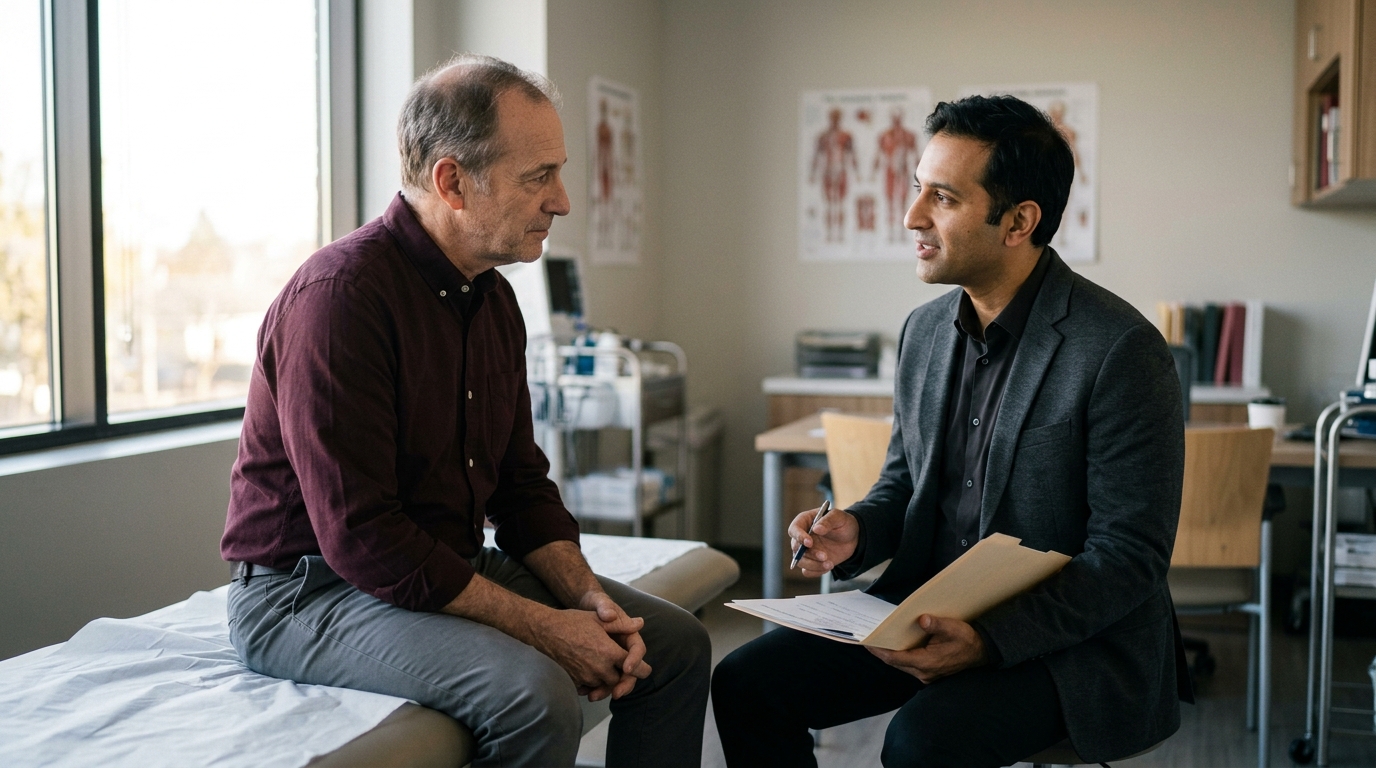

Most survivors expect normal energy levels to reflect intact hormone health, but cancer therapies often disrupt endocrine pathways independently from fertility functions.





Medical Disclaimer: This guide is for educational purposes only and does not constitute individual medical advice. Diagnosing and managing hormonal changes after cancer treatment requires a personalized evaluation by a qualified medical oncologist, urologist, or endocrinologist. Never start, alter, or discontinue any hormone therapy without direct supervision from your healthcare team.
A common assumption among cancer survivors is that successful treatment means the body immediately returns to its previous baseline. Another common belief is that any major cancer therapy permanently eliminates hormone production. Clinical research demonstrates that neither assumption is accurate. Endocrine changes following oncology care follow nuanced pathways. Some men maintain stable hormone levels, while others face delayed deficits that emerge months or years later.
Understanding how cancer therapies affect hormonal health requires a close look at biological mechanisms, specific treatment exposures, and clinical monitoring. This comprehensive guide reviews how surgery, chemotherapy, and radiation can influence testosterone pathways. It outlines how clinicians distinguish hormonal deficiency from fertility loss and explains how to approach long-term survivorship care.
A critical step in evaluating post-treatment health is separating testosterone production from sperm production. While both functions occur in the male gonads, they rely on entirely different cellular systems and control mechanisms. Conflating these two functions often creates confusion for survivors and their families.
Testosterone is synthesized by Leydig cells in the interstitial tissue of the testes. This process is stimulated primarily by luteinizing hormone, also known as LH, which is secreted by the pituitary gland. In contrast, sperm production occurs within the seminiferous tubules. This process is driven by germ cells and Sertoli cells under the stimulation of follicle-stimulating hormone, or FSH. Because these cellular populations have different vulnerabilities, cancer treatments can damage one system while leaving the other intact.
Having a normal total testosterone level does not guarantee preserved fertility. Conversely, severe impairment of sperm production does not mean that testosterone levels are deficient. For instance, germ cells are exceptionally sensitive to radiation and cytotoxic chemotherapy. Leydig cells are generally more resilient, though they can still suffer acute or permanent injury under higher treatment doses.
Pre-existing baseline function adds another layer of complexity. Research published in testicular cancer survivorship reviews shows that up to 50 percent of men with testicular cancer present with abnormal semen parameters before receiving any treatment. Up to 24 percent show complete absence of sperm, known as azoospermia, prior to therapy. These findings demonstrate that baseline health can be altered by the underlying disease itself before oncology interventions begin.
Recovery timelines also differ markedly between hormonal output and sperm production. Following platinum-based chemotherapy, approximately 20 percent of men experience azoospermia at one year. Some degree of spermatogenesis recovers in roughly 48 percent of men at two years and 80 percent at five years. These figures describe sperm recovery and must not be interpreted as testosterone recovery rates.
Recognizing this distinction is essential when considering medical therapies. Exogenous testosterone replacement therapy acts as a strong contraceptive by shutting down pituitary LH and FSH secretion. If a survivor with low testosterone receives testosterone replacement, his remaining sperm production will likely cease. Therefore, clinicians advise against testosterone replacement for any patient who desires future biological children until fertility goals and preservation options are fully addressed.
Cancer therapies can alter testosterone output through primary or secondary pathways. Primary gonadal dysfunction occurs when cancer treatments directly damage the testes and impair Leydig cell capacity. Secondary hypogonadism occurs when therapies affect the brain, specifically the hypothalamus or pituitary gland, blunting the hormonal signals that instruct the testes to make testosterone.
Unilateral orchiectomy is the standard surgical removal of one testicle for testicular cancer. Most men retain normal serum testosterone concentrations after undergoing a single orchiectomy because the remaining testicle compensates. However, long-term risk remains present. In a large surveillance cohort followed for a median of 11.4 years, 11 percent of patients managed with surveillance alone had total testosterone levels below 10 nmol/L.
Surgical procedures in the abdomen and pelvis can also affect reproductive function without altering testosterone levels. Retroperitoneal lymph node dissection, often abbreviated as RPLND, is performed to remove abdominal lymph nodes where cancer cells may spread. RPLND does not remove hormone-producing tissue, but it can affect the sympathetic nerves controlling ejaculation.
Survivorship data show that fertility rates reach approximately 62 percent following nerve-sparing RPLND, compared to 37 percent following non-nerve-sparing techniques. This difference relates to ejaculatory mechanics and retrograde ejaculation rather than Leydig cell failure. Keeping these distinctions clear prevents patients from attributing surgical nerve effects to hormonal deficiency.
Cytotoxic medications can cause direct, dose-dependent damage to testicular tissue. A comprehensive meta-analysis evaluating testicular cancer survivors revealed that men receiving conventional chemotherapy faced an odds ratio of 1.8 for developing testosterone deficiency compared to those managed with orchiectomy alone. For those receiving non-conventional chemotherapy, defined as more than four cycles or combined chemotherapy and radiation, the odds ratio rose to 3.1.
The Cancer Council Australia highlights specific risk factors for treatment-related Leydig cell impairment. These include regimens such as BEP (bleomycin, etoposide, and cisplatin), cumulative exposure exceeding four cycles of cisplatin, and combined radiation therapy. The risk of long-term deficiency is particularly elevated in patients who demonstrate an elevated LH level above 8 U/L after treatment.
Drug selection and cumulative dosage play significant roles in overall gonadal toxicity. Research indicates that carboplatin generally carries lower gonadal toxicity than cisplatin, and single-dose adjuvant regimens produce milder effects than multi-cycle therapeutic regimens. These patterns reflect general statistical associations. They do not predict the exact hormonal outcome for any single survivor, as individual susceptibility varies widely.
Radiation impacts hormonal pathways based on the anatomical target, total delivered dose, and shielding protocols. Infradiaphragmatic radiation therapy, which targets abdominal and pelvic lymph nodes, is associated with an odds ratio of 1.6 for testosterone deficiency compared to surgery alone. When gonadal shielding is applied during retroperitoneal radiation, scatter radiation to the remaining testicle is reduced, lowering the risk to Leydig cells.
Radiation delivered to the brain operates through an entirely different mechanism. Brain irradiation can damage the pituitary gland and hypothalamus. The Canadian Cancer Society notes that cranial radiation can suppress pituitary secretion of both LH and FSH.
This central suppression creates secondary hypogonadism, leading to reduced testosterone synthesis and diminished sperm production despite completely healthy testicular tissue. Recognizing the radiation field is critical for ordering the proper diagnostic lab tests. Direct testicular injury causes elevated LH levels, whereas central pituitary damage results in low or inappropriately normal LH levels.
Identifying low testosterone after cancer treatment requires a disciplined approach. Symptoms of hormone deficiency are broad and non-specific, frequently overlapping with other physical and psychological conditions. A survivor experiencing fatigue or low mood cannot assume testosterone deficiency is the culprit without rigorous diagnostic evaluation.
The 2022 testicular cancer survivorship review lists key clinical symptoms and physical signs that should prompt an endocrine assessment:
While these symptoms indicate a need for medical review, they do not confirm a hormone deficiency. Many survivors face long-term physical fatigue from cancer-related deconditioning, chronic inflammation, or psychological distress. Mood disorders, cardiovascular disease, thyroid dysfunction, and sleep disorders can present with identical patterns.
When a patient reports hallmark symptoms but morning blood testing reveals normal testosterone concentrations, clinicians must look for other causes. The survivorship review emphasizes evaluating alternative diagnoses, including cardiovascular complications, nutritional deficits, psychological adjustment challenges, or cancer recurrence. Jumping to hormone therapy based solely on subjective complaints without biochemical confirmation risks missing serious underlying medical conditions.
A precise laboratory evaluation requires measuring specific biomarkers under standardized conditions. Total testosterone alone does not provide a complete clinical picture. Interpreting hormone levels requires understanding binding proteins and central feedback signals. Learning about blood testing and biomarker evaluation helps survivors navigate their lab results.
Total testosterone measures all circulating testosterone in the bloodstream. Most circulating testosterone is bound tightly to sex hormone-binding globulin (SHBG) or bound loosely to albumin. Only a small fraction, typically one to two percent, remains unbound as free testosterone. Free testosterone and albumin-bound testosterone constitute bioavailable testosterone, which is readily accessible to bodily tissues.
Cancer treatments and metabolic changes can significantly alter SHBG levels, skewing total testosterone readings. For example, acute illness, liver changes, and alterations in thyroid function can shift SHBG concentrations up or down. If SHBG is unusually high, total testosterone may appear normal while free testosterone is clinically deficient. If SHBG is low, total testosterone may look depressed while free testosterone remains adequate.
The Endocrine Society clinical practice guideline mandates that diagnosis requires consistent signs and symptoms paired with unequivocally low serum testosterone concentrations. Testing must be performed on a fasting morning blood sample, collected between 7:00 AM and 10:00 AM. Because testosterone levels fluctuate daily, an initial low result must be confirmed with a repeat morning measurement before establishing a diagnosis.
Luteinizing hormone (LH) and follicle-stimulating hormone (FSH) are pituitary gonadotropins that reveal how the brain communicates with the testes. Measuring these hormones helps clinicians pinpoint the biological location of endocrine disruption:
Cancer Council Australia recommends monitoring testosterone, LH, and FSH as part of routine post-treatment reproductive follow-up. Combining gonadotropin measurements with morning testosterone allows clinicians to distinguish between testicular injury, central brain damage, or normal physiological function.
Clinicians may evaluate other biomarkers to rule out secondary conditions that suppress the hypothalamic-pituitary-gonadal axis:
Clinical guidance and evidence quality vary significantly across different cancer survivor groups. Guidelines tailored to adult germ-cell tumor survivors cannot be applied interchangeably to childhood cancer survivors or lymphoma patients. Understanding where research is robust and where data remain observational is essential for interpreting clinical recommendations.
The American Urological Association (AUA) provides clear guidance for adult germ-cell tumor survivors. The AUA recommends monitoring patients for clinical signs and symptoms of hypogonadism during routine survivorship care. If symptoms appear, the clinician should measure morning total testosterone and LH. This symptom-triggered framework focuses testing on patients experiencing clinical changes.
Cancer Council Australia provides a broader surveillance recommendation for post-pubertal boys and men. It recommends initiating reproductive follow-up 12 months after cancer therapy ends. Ongoing follow-up should be guided by original pathology, specific treatments received, and current symptoms, incorporating testosterone, LH, and FSH blood panels.
Guidance for childhood cancer survivors follows a separate framework established by organizations like the Children’s Oncology Group (COG). The COG guidelines note that overt testosterone deficiency requiring lifelong replacement is rare after treatment with alkylating agents alone. However, the COG recommends formal endocrine referral for childhood survivors who present with low morning testosterone or show delayed pubertal progression by age 14.
The COG also advises periodic long-term re-evaluation for adult males who maintain low-normal testosterone levels as they age. Because aging causes a natural decline in testosterone, men with baseline subclinical damage from childhood treatments may develop overt deficiency earlier in life. These population-specific differences illustrate why survivors need individualized follow-up plans tailored to their age, cancer type, and exposure history.
Testosterone plays an integral role in male metabolic and cardiovascular regulation. Survivors who develop long-term hormone deficiency face risks beyond sexual dysfunction and fatigue. Hypogonadism is strongly linked to adverse changes in lipid metabolism, insulin sensitivity, visceral fat accumulation, and arterial vascular health.
The AUA testicular cancer guidelines emphasize that survivorship care must address late toxicities, particularly for men who received chemotherapy, radiotherapy, or combined modalities. These treatments can accelerate cardiovascular and metabolic risk factors. Long-term survivors have higher rates of hypertension, hyperlipidemia, metabolic syndrome, and early coronary artery disease compared to age-matched controls.
Low testosterone can exacerbate these metabolic complications. Testosterone deficiency promotes the accumulation of visceral abdominal fat, which increases systemic inflammatory cytokines and promotes insulin resistance. Over time, this state increases the likelihood of developing type 2 diabetes and endothelial dysfunction.
Survivorship clinics emphasize comprehensive primary care monitoring for this reason. A structured follow-up program should track:
Integrating hormone monitoring into broader cardiovascular surveillance ensures that clinicians address the total health of the survivor. Treating metabolic risks through lifestyle adjustments, exercise, dietary support, and medical management reduces the long-term burden of cancer therapy. Survivors can learn more about general hormone health by reading about testosterone fundamentals.
Balancing hormone replacement therapy with fertility goals is one of the most complex clinical challenges in oncology survivorship. The American Society of Clinical Oncology (ASCO) updated its clinical practice guidelines to emphasize that fertility preservation must be discussed at initial diagnosis and re-evaluated throughout survivorship.
Sperm cryopreservation remains the gold standard for male fertility preservation prior to undergoing gonadotoxic therapy. ASCO and urological guidelines advise offering sperm banking to all men presenting with testicular cancer or other malignancies requiring chemotherapy, radiotherapy, or RPLND. This includes adolescents and men who are undecided about their future family plans.
Clinicians often discuss banking sperm prior to orchiectomy. While pre-orchiectomy banking can secure viable sperm in men who might become azoospermic post-surgery, it must be balanced against surgical scheduling, financial costs, and individual clinical necessity. If pre-treatment banking was not performed, post-treatment cryopreservation should be explored once the patient reaches remission.
When a survivor is diagnosed with both symptomatic hypogonadism and persistent infertility, the treatment plan must be carefully sequenced. The testicular cancer survivorship review clearly outlines that exogenous testosterone replacement therapy should never be prescribed to men actively seeking biological children. Exogenous testosterone creates negative feedback on the pituitary gland, halting LH and FSH release and shutting down testicular spermatogenesis.
For men with confirmed testosterone deficiency who wish to preserve or restore fertility, clinicians consider alternative strategies:
When fertility is no longer desired and a patient meets strict diagnostic criteria, testosterone replacement therapy may be considered. Hormone therapy aims to restore circulating testosterone to the physiological range, improving bone density, lean body mass, energy, and sexual function. Once replacement begins, clinicians must monitor both symptomatic improvements and biochemical markers, ensuring hematocrit, prostate health, and cardiovascular markers remain within safe parameters.
Open communication with healthcare providers ensures that potential hormone deficiencies are recognized early and managed safely. Survivors should feel empowered to bring specific, structured questions to their follow-up appointments.
Consider discussing these practical questions with your oncologist, urologist, or endocrinologist:
Understanding the biological causes of low testosterone allows survivors to navigate these conversations with confidence. Sharing details about your energy, recovery, sexual function and energy levels, and cognitive focus provides your care team with the clinical data needed to optimize your long-term health.
Navigating hormonal recovery after cancer treatment requires patience, precise testing, and open collaboration with an experienced medical team.
Testostra explains testosterone biology, testing, symptoms, lifestyle factors and TRT with careful sourcing and clear clinical limits.



Send us a question, research idea or topic suggestion. Reader questions help shape future Testostra content.
Contact Testostra


































































































